A joint research study conducted by Punjabi University, Patiala, and the Post Graduate Institute of Medical Education and Research (PGIMER), Chandigarh, has resulted in the development of artificial intelligence (AI) based methods to diagnose Autoimmune Blistering Diseases (AIBDs). These rare, complex skin disorders traditionally require expensive and specialized diagnostic tests, such as Direct Immunofluorescence (DIF), which are often unavailable in resource-limited settings. By creating a clinically validated dataset of patient skin lesion images, the research team trained multiple AI models that demonstrated superior accuracy over human dermatologists in classifying specific AIBD subtypes. This innovation aims to provide primary and rural healthcare clinicians with an accessible, cost-effective tool to diagnose rare diseases early and accurately.
Understanding Autoimmune Blistering Diseases (AIBDs)
Pathophysiology and Nature of AIBDs
AIBDs are a group of rare, life-threatening systemic disorders characterized by the formation of blisters on the skin and mucous membranes. The condition occurs when the body’s immune system mistakenly produces autoantibodies that target structural proteins responsible for maintaining skin integrity. These proteins hold epidermal cells together or bind the epidermis to the underlying dermis.
Major Subtypes of AIBDs
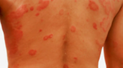
The diseases are broadly classified based on the layer of skin where the blistering occurs:
- Intraepidermal Blistering (Pemphigus Group): The autoantibodies attack proteins like desmogleins within the upper layer of the skin (epidermis). This causes cells to separate, a process known as acantholysis. Pemphigus vulgaris and Pemphigus foliaceus are the primary forms.
- Subepidermal Blistering (Pemphigoid Group): The immune attack targets the basement membrane zone that connects the epidermis to the dermis. Bullous pemphigoid is the most common subtype, typically affecting elderly populations, along with Mucous Membrane Pemphigoid and Epidermolysis Bullosa Acquisita.
Traditional Diagnostic Challenges
Limitations of Direct Immunofluorescence (DIF)
The gold standard for diagnosing AIBDs is Direct Immunofluorescence (DIF) microscopy performed on a skin biopsy. This process requires specialized laboratory infrastructure, highly trained pathologists, and expensive chemical reagents.
Accessibility and Cost Barriers
DIF testing facilities are heavily concentrated in tertiary care medical centers and urban hospitals. Patients in rural or primary healthcare settings face delayed diagnoses due to the high cost of transportation, biopsy procedures, and laboratory fees. Misdiagnosis at early stages often leads to inappropriate treatments and worsening of the clinical condition.
The AI-Based Diagnostic Solution
Dataset Creation and Clinical Validation
The collaboration between Punjabi University and PGIMER involved compiling a comprehensive, expert-annotated dataset of clinical images showcasing various stages and subtypes of AIBD skin lesions. Dermatologists and pathologists validated each image to ensure the ground-truth data was highly accurate before introducing it to the machine learning pipeline.
Model Training and Performance
The researchers deployed Deep Learning (DL) architectures, specifically Convolutional Neural Networks (CNNs), which excel at computer vision tasks like medical image classification. When tested, these AI models achieved classification accuracy rates that outperformed practicing dermatologists, successfully distinguishing between look-alike blistering subtypes based on subtle visual patterns in the lesions.
Deployment in Primary Healthcare
The lightweight nature of the finalized AI algorithms allows them to be integrated into tele-dermatology platforms or mobile applications. Healthcare workers in rural clinics can capture an image of a patient’s skin blister using a standard smartphone, upload it to the AI system, and receive an immediate diagnostic probability score to guide referral or initial management.
| Parameter | Traditional Diagnosis (DIF) | AI-Assisted Diagnosis |
| Primary Requirement | Skin biopsy and specialized microscopy | Digital image of the skin lesion |
| Infrastructure Needed | Tertiary care pathology labs | Smartphone / Tele-medicine portal |
| Time for Results | Several days to weeks | Real-time / Immediate analysis |
| Cost to Patient | High (Biopsy + Lab processing) | Nominal / Low-cost digital check |
| Skill Requirement | Expert Dermatopathologist | Primary Healthcare Worker |
IASPOINT Booster Facts for UPSC
- Epidermis vs. Dermis: The skin consists of three main layers: the epidermis (outermost waterproof barrier), the dermis (contains tough connective tissue, hair follicles, and sweat glands), and the hypodermis (deeper subcutaneous fat and tissue).
- Autoantibodies: These are antibodies produced by the immune system that mistakenly target and react with a person’s own tissues or organs, failing to distinguish between self and non-self proteins.
- Convolutional Neural Networks (CNNs): A class of artificial neural networks most commonly applied to analyzing visual imagery. They use a mathematical operation called convolution to process pixel data and detect edges, textures, and complex shapes.
- National Policy for Rare Diseases (NPRD), 2021: India’s policy classifies rare diseases into three groups based on treatment cost and availability. It provides financial support of up to ₹50 lakhs for patients suffering from any category of rare diseases for treatment at designated Center of Excellences (CoEs).
- Ayushman Bharat Digital Mission (ABDM): This initiative aims to develop the backbone necessary to support the integrated digital health infrastructure of India. AI diagnostic tools can easily link with ABDM-compliant registries to streamline rural specialist consultations.