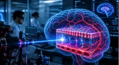
MIT researchers announced a bioimaging technique on 27–28 April 2026 that uses a self-organising “pencil beam” laser for 3D imaging of the human blood-brain barrier. The method achieves imaging speeds about 25 times faster than existing gold-standard techniques while maintaining comparable resolution.
Laser Beam Formation
The “pencil beam” forms when laser light enters a multimode optical fibre at a precise zero-degree angle and at a power level sufficient to interact with the fibre’s glass. The beam develops from chaotic laser light under specific physical conditions.
Imaging Applications
The technique allows researchers to observe individual cells absorbing drugs in real time. It can track diverse compounds and molecular targets across engineered tissue models, which makes it useful for studies of neurological diseases such as Alzheimer’s disease and amyotrophic lateral sclerosis (ALS).
Research Publication
The findings appeared in the international journal Nature Methods on 27 April 2026. The MIT team included senior author Sixian You and lead graduate student Honghao Cao.
Technical Context
A multimode optical fibre carries multiple light paths through a single fibre core. The blood-brain barrier is a selective barrier formed by brain blood vessels that regulates the movement of substances between the bloodstream and the brain.
Last Modified: April 29, 2026