
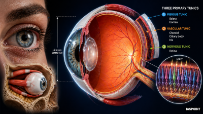
The human eye is a complex sensory organ specialized for photoreception. It is nearly spherical in shape, approximately 2.4 cm in diameter, and is housed within the bony orbits of the skull. The eye consists of three primary layers or “tunics.”
1. The External Layer: Fibrous Tunic
This is the outermost, protective layer of the eyeball, composed of dense connective tissue.
- Sclera: The “white” of the eye. It provides structural integrity and serves as an attachment point for the extrinsic eye muscles that move the eyeball.
- Cornea: The anterior, transparent portion of the sclera. It is avascular (lacks blood vessels) and derives nutrition from the aqueous humor. It is the primary structure responsible for refracting (bending) light entering the eye.
2. The Middle Layer: Vascular Tunic (Uvea)
This layer is highly vascularized and contains pigments that prevent light reflection within the eye.
- Choroid: A thin, bluish layer containing numerous blood vessels. It provides nourishment to the retina.
- Ciliary Body: The anterior thickening of the choroid. It contains ciliary muscles that alter the shape of the lens for near and far vision (Accommodation).
- Iris: The visible colored portion of the eye. It acts as a diaphragm with a central opening called the Pupil. The iris regulates the amount of light entering the eye by adjusting the diameter of the pupil.
3. The Inner Layer: Neural Tunic (Retina)
The retina is the light-sensitive innermost layer where the process of visual transduction occurs.
- Cellular Organization: The retina consists of three layers of neurons (from inside to outside):
- Ganglion Cells (Axons form the optic nerve)
- Bipolar Cells
- Photoreceptor Cells (Rods and Cones)
- Optic Disc (Blind Spot): The area where the optic nerve exits the eyeball. It contains no photoreceptors; hence, no image is formed here.
- Macula Lutea: A yellowish central area of the retina.
- Fovea Centralis: A small pit in the center of the macula containing only densely packed cones. It is the region of highest visual acuity (sharpness).
Internal Components and Media
The interior of the eye is divided into two distinct chambers filled with fluid, separated by the lens.
| Component | Description | Function |
| Lens | Transparent, crystalline, biconvex structure. | Focuses light onto the retina; held by suspensory ligaments. |
| Aqueous Humor | Thin, watery fluid in the anterior chamber (between cornea and lens). | Maintains intraocular pressure and nourishes the cornea/lens. |
| Vitreous Humor | Transparent gel in the posterior chamber (between lens and retina). | Maintains the spherical shape of the eyeball and supports the retina. |
Mechanism of Vision: Transduction Process
- Light passes through the Cornea and Lens, focusing on the Retina.
- Light induces the dissociation of the photopigment Rhodopsin into Opsin (protein) and Retinal (an aldehyde of Vitamin A).
- This dissociation changes the membrane permeability of the photoreceptor cells, generating an action potential.
- The impulse is transmitted through Bipolar cells to Ganglion cells.
- The Optic Nerve carries these impulses to the visual cortex of the brain for image processing.
Key Facts for UPSC Prelims
- Corneal Transplant: The cornea is the only tissue in the human body that can be transplanted without the need for tissue matching because it is avascular and does not trigger an immune response.
- Night Blindness (Nyctalopia): Caused by a deficiency of Vitamin A, which leads to a lack of Rhodopsin in the rods.
- Glaucoma: A condition caused by increased intraocular pressure due to the blockage of aqueous humor drainage.
- Cataract: Characterized by the lens becoming opaque or cloudy, usually due to aging, leading to impaired vision.