
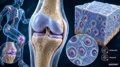
Cartilage is a specialized, tough, yet semi-flexible supporting connective tissue. It is characterized by an extracellular matrix that is solid and pliable, allowing it to resist compression while remaining elastic. Unlike bone, cartilage is avascular (lacks blood vessels) and aneural (lacks nerves), which significantly impacts its ability to heal after injury.
Composition of Cartilage
The structural integrity of cartilage is maintained by specific cellular and chemical components.
1. Specialized Cells
- Chondroblasts: Immature cells that actively secrete the extracellular matrix.
- Chondrocytes: Mature cartilage cells located in small fluid-filled cavities called lacunae. They maintain the health of the cartilage matrix.
2. Extracellular Matrix (ECM)
- Ground Substance: Rich in Chondroitin salts and proteins, which provide the tissue with its characteristic resilience.
- Fibers: Contains varying proportions of collagen and elastic fibers depending on the specific type of cartilage.
Classification of Cartilage
Cartilage is categorized into three types based on the nature of the matrix and the predominant fiber type.
| Type | Characteristics | Key Locations |
| Hyaline Cartilage | Most abundant; bluish-white and “glassy”; rich in fine collagen fibers. | Articular surfaces of long bones, nose, larynx, trachea, and bronchial tubes. |
| Elastic Cartilage | Yellowish; rich in elastic fibers; maintains shape after repeated bending. | External ear (pinna), Eustachian tubes, and the Epiglottis. |
| Fibrocartilage | Strongest type; contains thick bundles of Type I collagen; high tensile strength. | Intervertebral discs, pubic symphysis, and the discs of the knee joint (menisci). |
Vital Functions of Cartilage
- Support and Flexibility: Provides a framework for structures like the nose and ears while maintaining their shape.
- Shock Absorption: Acts as a cushion between bones in joints, preventing wear and tear during movement.
- Fetal Skeleton: Serves as the template for the human skeleton during embryonic development. Most of this cartilage is later replaced by bone through Endochondral Ossification.
- Smooth Movement: Lines the ends of bones (articular cartilage) to reduce friction in synovial joints.
Key Differences: Cartilage vs. Bone
| Feature | Cartilage | Bone |
| Matrix | Semi-solid, pliable, non-calcified. | Hard, rigid, calcified (Calcium salts). |
| Blood Supply | Avascular (receives nutrients via diffusion). | Highly vascular. |
| Nerves | Absent. | Present. |
| Growth | Appositional and Interstitial. | Appositional only. |
| Healing | Very slow/limited. | Fast and efficient. |
Vital Facts for UPSC Prelims
- Perichondrium: A layer of dense irregular connective tissue that surrounds most cartilage. It contains the blood vessels that supply nutrients to the cartilage cells via diffusion. Note: Fibrocartilage and articular cartilage lack a perichondrium.
- Diffusion Limit: Because cartilage is avascular, it has a limited thickness. If the tissue becomes too thick, the chondrocytes in the center cannot receive enough oxygen or nutrients and will die.
- Costal Cartilage: The specific hyaline cartilage that connects the ribs to the sternum, allowing the chest cavity to expand during breathing.
- Calcification in Aging: As humans age, some hyaline cartilage may undergo calcification, losing its flexibility and potentially causing joint pain (Osteoarthritis).
- Ear Piercing Trivia: The pain felt during a “cartilage piercing” is actually due to the trauma of the surrounding skin and perichondrium, as the cartilage itself lacks nerves.