
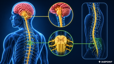
The spinal cord is a long, thin, tubular bundle of nervous tissue and support cells that extends from the Medulla Oblongata in the brainstem down to the lumbar region of the vertebral column. It serves as the primary conduit for signals between the brain and the rest of the body.
Structural Anatomy of the Spinal Cord
The spinal cord is housed within the vertebral canal formed by the vertebral column.
- Conus Medullaris: The tapered, lower end of the spinal cord, usually terminating near the first or second lumbar vertebra (L1 or L2) in adults.
- Cauda Equina: A bundle of spinal nerves and nerve rootlets at the base of the spinal cord, resembling a “horse’s tail,” which continues through the lower vertebral canal.
- Filum Terminale: A delicate strand of fibrous tissue that extends from the conus medullaris to the coccyx, providing longitudinal support.
Meningeal Protection and CSF
Like the brain, the spinal cord is protected by three layers of meninges:
- Dura Mater: The outermost, tough protective layer.
- Arachnoid Mater: The middle layer with web-like structures.
- Pia Mater: The innermost layer, closely adhering to the cord.
- Epidural Space: A space between the dura mater and the vertebral wall containing fat and small blood vessels. This is the site for “epidural” anesthesia.
- Cerebrospinal Fluid (CSF): Found in the Central Canal (a small hole in the center of the cord) and the subarachnoid space, providing mechanical cushioning.
Internal Composition: Gray and White Matter
The arrangement of tissue in the spinal cord is the inverse of the brain’s arrangement.
- Gray Matter (Inner): Shaped like a butterfly or the letter ‘H’ in cross-section. It contains neuronal cell bodies and interneurons.
- Dorsal Horn: Receives sensory information from the body (Afferent).
- Ventral Horn: Contains motor neurons that exit to muscles (Efferent).
- White Matter (Outer): Surrounds the gray matter and consists of bundles of myelinated axons called Tracts.
- Ascending Tracts: Carry sensory information upward to the brain.
- Descending Tracts: Carry motor commands downward from the brain.
Spinal Nerves
There are 31 pairs of spinal nerves that emerge from the spinal cord, categorized by the region of the vertebral column:
- Cervical (C1–C8): 8 pairs (Neck region).
- Thoracic (T1–T12): 12 pairs (Chest region).
- Lumbar (L1–L5): 5 pairs (Lower back).
- Sacral (S1–S5): 5 pairs (Sacrum).
- Coccygeal (Co1): 1 pair (Tailbone).
Physiological Functions
The spinal cord performs two critical roles in the nervous system:
- Signal Transduction: It acts as a bridge for sensory impulses traveling to the brain and motor impulses traveling to the periphery.
- Reflex Integration: It processes Reflex Actions independently of the brain. The spinal cord can receive a sensory signal and immediately send a motor command (e.g., pulling a hand away from a hot surface) before the brain even perceives the pain.
Reflex Arc Components
| Component | Function |
| Receptor | Detects the stimulus (e.g., heat, sharp prick). |
| Sensory Neuron | Transmits the impulse to the dorsal horn of the spinal cord. |
| Interneuron | Processes the signal within the gray matter (skips the brain for speed). |
| Motor Neuron | Transmits the command from the ventral horn to the muscle. |
| Effector | The muscle or gland that carries out the response. |
UPSC Prelims Fact File
- Foramen Magnum: The large opening at the base of the skull where the medulla oblongata connects to the spinal cord.
- Lumbar Puncture (Spinal Tap): A medical procedure where CSF is collected from the subarachnoid space, usually between L3 and L4 or L4 and L5, to avoid damaging the spinal cord itself.
- Meningitis: Inflammation of the meninges surrounding the brain and spinal cord, which can be bacterial or viral and is a significant public health topic.
- Reflex Centers: While the spinal cord handles simple skeletal reflexes, vital autonomic reflexes (respiration, heart rate) are handled by the Medulla Oblongata in the brainstem