
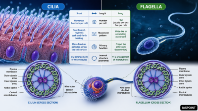
Cilia and flagella are hair-like outgrowths of the cell membrane. They are microtubule-based organelles responsible for locomotion and the movement of fluids or particles across the cell surface. While they share a similar internal structure, they differ in length, number per cell, and beating patterns.
Structural Architecture
Both cilia and flagella emerge from a centriole-like structure called the basal body.
The Axoneme and “9+2” Array
- Core Structure: The internal core is called the axoneme.
- Microtubule Arrangement: It consists of nine doublets of peripheral microtubules surrounding a pair of central microtubules. This is famously known as the 9+2 arrangement.
- Central Sheath: The central tubules are enclosed by a sheath and connected to one of the tubules of each peripheral doublet by a radial spoke.
- Linkers: Peripheral doublets are interconnected by linkers called nexin and possess motor proteins called dynein arms that facilitate movement.
Functional Comparison: Cilia vs. Flagella
| Feature | Cilia | Flagella |
| Length | Short (5–10 micrometers) | Long (up to 150 micrometers) |
| Number | Numerous (hundreds per cell) | Few (usually 1 to 4) |
| Motion | Oar-like, coordinated rhythm | Undulatory, whip-like independent motion |
| Function | Movement of cell or surrounding fluid | Primarily cell locomotion |
| Occurrence | Eukaryotes only | Prokaryotes and Eukaryotes |
Diverse Roles in the Human Body
Ciliary Functions
- Respiratory Tract: Ciliated epithelium lines the trachea and bronchi to sweep mucus and trapped dust particles upward toward the throat (the “mucociliary escalator”).
- Fallopian Tubes: Cilia help in the movement of the ovum toward the uterus.
- Non-motile (Primary) Cilia: Almost all mammalian cells have a single “primary cilium” that acts as a sensory antenna for chemical and mechanical signals.
Flagellar Functions
- Spermatozoa: The flagellum provides the motive force for the sperm cell to swim through the female reproductive tract.
Prokaryotic vs. Eukaryotic Flagella
It is a common point of confusion for UPSC aspirants to assume all flagella are the same. They are evolutionarily and structurally distinct.
- Eukaryotic Flagella: Composed of tubulin protein, move in a whip-like fashion, and are powered by ATP using a sliding microtubule mechanism.
- Prokaryotic Flagella: Composed of flagellin protein, move in a rotary (spinning) motion like a propeller, and are powered by a proton gradient (proton motive force).
Clinical Conditions: Ciliopathies
Defects in the structure or function of cilia lead to various genetic disorders:
- Kartagener’s Syndrome: A condition where dynein arms are absent, leading to immobile cilia. Symptoms include chronic respiratory infections and male infertility.
- Situs Inversus: Functional cilia are required during embryonic development to determine the left-right symmetry of organs. Defective cilia can result in organs being mirrored (e.g., heart on the right side).
Facts for UPSC Prelims
- Basal Body: The basal body from which cilia and flagella emerge has a 9+0 arrangement, identical to a centriole.
- Energy Source: The movement of eukaryotic cilia and flagella is an active process requiring ATP.
- Discovery: Antonie van Leeuwenhoek was the first to observe flagella in the 17th century.
- Protists: Organisms like Paramecium use cilia for both locomotion and for steering food into their gullet, whereas Euglena uses a flagellum for movement.