
A reflex action is an extremely rapid, involuntary, and predictable response to a peripheral nervous stimulation that occurs without conscious thought. These actions are governed primarily by the spinal cord, though the brain may receive the sensory information after the action has already been initiated. Reflexes are vital survival mechanisms designed to protect the body from immediate physical harm.
The Reflex Arc: Pathway of Nerve Impulses
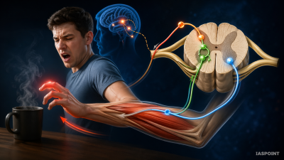
The anatomical pathway that controls a reflex action is called the Reflex Arc. It involves a specific circuit of nervous tissue that bypasses the higher processing centers of the brain for speed.
Components of a Reflex Arc
A standard reflex arc consists of five essential components:
- Receptor: Specialized cells or organs (like the skin or retina) that detect a stimulus (e.g., heat, pressure, or a sharp object).
- Sensory (Afferent) Neuron: Conducts the nerve impulse from the receptor toward the Central Nervous System (CNS), specifically entering the spinal cord through the dorsal root.
- Interneuron (Association Neuron): Located within the grey matter of the spinal cord, it processes the incoming signal and relays it directly to a motor neuron.
- Motor (Efferent) Neuron: Carries the response signal from the spinal cord to the effector organ via the ventral root.
- Effector: The muscle or gland that executes the final response (e.g., a muscle contracting to pull a hand away).
Types of Reflexes
Reflexes are classified based on their complexity and the nature of their development.
1. Simple (Unconditioned) Reflexes
These are inborn, hereditary responses that do not require previous learning or experience.
- Examples: Knee-jerk reflex, blinking of eyes when an object approaches, and the withdrawal reflex from a hot surface.
2. Acquired (Conditioned) Reflexes
These are developed through experience, training, or repetitive learning.
- Examples: Salivating at the smell of favorite food (Ivan Pavlov’s experiment), typing on a keyboard without looking, or braking a vehicle suddenly in an emergency.
Classification based on Synaptic Involvement
- Monosynaptic Reflex: Involves only one synapse between the sensory and motor neuron (no interneuron). The Knee-jerk (Patellar) reflex is the most common example.
- Polysynaptic Reflex: Involves one or more interneurons between the sensory and motor neurons. Most protective reflexes, like the withdrawal reflex, are polysynaptic.
Functional Significance of Reflex Actions
- Speed: By bypassing the brain’s decision-making process, reflexes reduce the “lag time” between stimulus and response, preventing tissue damage.
- Automation: They manage routine functions (like pupil constriction in bright light) without exhausting the brain’s cognitive resources.
- Diagnostic Tool: Clinicians test reflexes (like the Achilles reflex or Babinski sign) to assess the health of specific segments of the spinal cord and peripheral nerves.
Comparison: Reflex Action vs. Voluntary Action
| Feature | Reflex Action | Voluntary Action |
| Control Center | Mainly Spinal Cord | Cerebrum (Forebrain) |
| Willpower | Involuntary | Under conscious control |
| Speed | Very Rapid | Relatively Slower |
| Path | Reflex Arc | Complex neural pathways |
| Purpose | Immediate protection | Goal-oriented tasks |
Key Facts for UPSC Prelims
- The Knee-Jerk Reflex: Specifically tests the L2 through L4 segments of the spinal cord.
- Pupillary Light Reflex: An autonomic reflex where the pupil constricts in response to light to protect the retina; it is controlled by the midbrain.
- Dorsal vs. Ventral Roots: Sensory information always enters the spinal cord through the dorsal (posterior) root, while motor commands always exit through the ventral (anterior) root.
- Grey Matter Involvement: The integration of the reflex arc occurs in the H-shaped grey matter of the spinal cord.
- Proprioception: Specialized internal reflexes that inform the brain about the position and movement of body parts, essential for maintaining balance.