
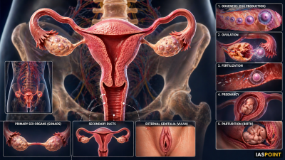
The female reproductive system is designed to perform complex functions including oogenesis (egg production), ovulation, fertilization, pregnancy, and parturition (birth). It is located in the pelvic region and consists of primary sex organs, secondary ducts, and external genitalia.
Primary Sex Organ: The Ovaries
The ovaries are the primary female sex organs that produce the female gamete (ovum) and several steroid hormones (estrogen and progesterone).
- Location: One ovary is located on each side of the lower abdomen, connected to the pelvic wall and uterus by ligaments.
- Structure: Each ovary is covered by a thin epithelium which encloses the ovarian stroma.
- Stroma Divisions: The stroma is divided into two zones: a peripheral cortex and an inner medulla.
- Ovarian Follicles: The cortex contains follicles at various stages of development. A mature follicle is known as a Graafian follicle.
The Female Accessory Ducts
The duct system consists of the Fallopian tubes, the uterus, and the vagina.
- Fallopian Tubes (Oviducts): Each tube is about 10–12 cm long.
- Infundibulum: The funnel-shaped part closer to the ovary. It possesses finger-like projections called fimbriae which help in collecting the ovum after ovulation.
- Ampulla: The wider, middle part of the oviduct where fertilization typically occurs.
- Isthmus: The narrow last part that joins the uterus.
- Uterus (Womb): An inverted pear-shaped muscular organ. It is supported by ligaments attached to the pelvic wall.
- Vagina: A muscular tube that opens to the exterior, serving as the birth canal and the receptacle for sperm.
Layers of the Uterine Wall
The wall of the uterus has three distinct layers of tissue:
| Layer | Type | Function |
| Perimetrium | External | Thin membranous covering. |
| Myometrium | Middle | Thick layer of smooth muscle; exhibits strong contractions during delivery. |
| Endometrium | Internal | Glandular layer that lines the uterine cavity; undergoes cyclic changes during the menstrual cycle. |
External Genitalia (Vulva)
The female external genitalia include:
- Mons Pubis: A cushion of fatty tissue covered by skin and pubic hair.
- Labia Majora: Fleshy folds of tissue that surround the vaginal opening.
- Labia Minora: Paired folds of tissue under the labia majora.
- Hymen: A membrane partially covering the opening of the vagina.
- Clitoris: A tiny finger-like structure at the upper junction of the labia minora; it is homologous to the male penis.
Process of Oogenesis
Oogenesis is the process of formation of a mature female gamete. Unlike spermatogenesis, it is initiated during the embryonic development stage.
- Multiplication Phase: Millions of gamete mother cells (oogonia) are formed within each fetal ovary; no more oogonia are formed or added after birth.
- Prophase-I Arrest: Oogonia enter meiosis-I but get temporarily arrested at the prophase stage, called primary oocytes.
- Follicular Atresia: A large number of primary follicles degenerate from birth to puberty. Only about 60,000–80,000 primary follicles are left in each ovary at puberty.
- Maturation: At puberty, a primary oocyte completes its first meiotic division (unequal), resulting in a large haploid secondary oocyte and a tiny first polar body.
- Ovulation: The Graafian follicle ruptures to release the secondary oocyte from the ovary.
The Menstrual Cycle
The reproductive cycle in female primates (monkeys, apes, and humans) is called the menstrual cycle.
- Menarche: The first menstruation begins at puberty.
- Menstrual Phase: Lasts 3–5 days; results in the breakdown of the endometrial lining if fertilization does not occur.
- Follicular (Proliferative) Phase: Primary follicles grow into Graafian follicles; the endometrium regenerates through proliferation.
- Ovulatory Phase: Rapid secretion of LH (LH surge) induces the rupture of the Graafian follicle and release of the ovum (Day 14).
- Luteal (Secretory) Phase: The remaining parts of the Graafian follicle transform into the Corpus Luteum, which secretes large amounts of progesterone to maintain the endometrium for potential pregnancy.
- Menopause: The cessation of menstrual cycles, usually occurring around age 50.
Mammary Glands
Functional mammary glands are characteristic of all female mammals. They are paired structures (breasts) containing glandular tissue and fat.
- Glandular Tissue: Divided into 15–20 mammary lobes.
- Alveoli: Clusters of cells within lobes that secrete milk, which is stored in the cavities (lumens) of alveoli.
- Duct System: Alveoli open into mammary tubules, which join to form a mammary duct. Several ducts join to form a wider mammary ampulla, connected to the lactiferous duct through which milk is sucked out.
Fact File for UPSC Prelims
- Corpus Luteum: Acting as a temporary endocrine gland, it is essential for maintaining the endometrium via progesterone. If fertilization fails, it transforms into the Corpus Albicans.
- Fertilization Site: Specifically occurs at the ampullary-isthmic junction of the Fallopian tube.
- Colostrum: The milk produced during the initial few days of lactation; it contains several antibodies (IgA) essential to develop resistance in newborns.
- HCG (Human Chorionic Gonadotropin): Produced only during pregnancy; its presence in urine is the basis for most pregnancy tests.