
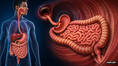
The alimentary canal, also known as the gastrointestinal (GI) tract, is a continuous muscular tube extending from the mouth to the anus. In a typical adult, it measures approximately 9 meters in length. It is the primary site for ingestion, digestion, absorption, and egestion.
Anatomical Structure: The Four Layers (Histology)
From the esophagus to the anal canal, the wall of the alimentary canal possesses four distinct tissue layers. Understanding these is crucial for Prelims as they relate to specific physiological functions:
- Serosa: The outermost layer made of thin mesothelium with some connective tissue.
- Muscularis: Formed by smooth muscles usually arranged into an inner circular and an outer longitudinal layer. An oblique muscle layer is uniquely present in the stomach.
- Sub-mucosa: Formed of loose connective tissues containing nerves, blood, and lymph vessels. In the duodenum, glands are also present in this layer.
- Mucosa: The innermost lining. It forms irregular folds (rugae) in the stomach and small finger-like foldings (villi) in the small intestine to increase surface area.
Organs of the Alimentary Canal
The Buccal Cavity and Pharynx
- Teeth: Humans exhibit thecodont (embedded in jaw bone), diphyodont (two sets of teeth: deciduous and permanent), and heterodont dentition.
- Dental Formula: The adult human dental formula is 2123/2123 (Incisors, Canines, Premolars, Molars).
- Tongue: A freely movable muscular organ attached to the floor of the oral cavity by the frenulum.
- Pharynx: A common passage for food and air. The epiglottis, a cartilaginous flap, prevents food from entering the glottis (windpipe opening) during swallowing.
The Esophagus and Stomach
- Esophagus: A thin, long tube which passes through the neck, thorax, and diaphragm. It conducts food via peristalsis.
- Stomach: A J-shaped muscular bag located in the upper left portion of the abdominal cavity.
- Cardiac: Region where the esophagus opens.
- Fundic: The superior dome-shaped region.
- Body: Main central region.
- Pyloric: Portion which opens into the first part of the small intestine.
- Sphincter: The gastro-esophageal sphincter controls entry from the esophagus, while the pyloric sphincter guards the exit to the duodenum.
The Small Intestine
The small intestine is the longest part of the alimentary canal and the principal site of nutrient absorption. It is divided into three regions:
- Duodenum: C-shaped (or U-shaped) shortest part; receives bile and pancreatic juice.
- Jejunum: Long, coiled middle portion.
- Ileum: Highly coiled region that opens into the large intestine.
The Large Intestine
Consists of the Caecum, Colon, and Rectum.
- Caecum: A small blind sac which hosts some symbiotic micro-organisms. The vermi-form appendix, a vestigial organ, arises from it.
- Colon: Divided into four parts—ascending, transverse, descending, and sigmoid colon.
- Rectum: Opens out through the anus.
Comparative Summary of Digestive Processes
| Organ | Primary Secretion/Feature | Major Function |
| Mouth | Salivary Amylase (Ptyalin) | Carbohydrate digestion starts (30% starch). |
| Stomach | HCl, Pepsin, Mucus | Protein digestion; kills pathogens; formation of chyme. |
| Duodenum | Bile (Liver) & Pancreatic Juice | Emulsification of fats; breakdown of all macromolecules. |
| Ileum | Succus Entericus (Intestinal Juice) | Final enzymatic breakdown into absorbable units. |
| Large Intestine | Mucus | Water and mineral absorption; feces formation. |
Critical UPSC Trivia: Did You Know?
- Brunner’s Glands: These are located specifically in the sub-mucosa of the duodenum and secrete alkaline mucus to protect the intestine from stomach acid.
- Peyer’s Patches: Lymphoid tissue found in the ileum; they are part of the gut-associated lymphoid tissue (GALT) and monitor bacterial populations.
- Goblet Cells: Special cells in the mucosal epithelium that secrete mucus for lubrication.
- Villi and Lacteals: Villi are supplied with a network of capillaries and a large lymph vessel called a lacteal, which is essential for the absorption of fats.
- Sphincter of Oddi: The hepato-pancreatic duct (carrying both bile and pancreatic juice) is guarded by this sphincter before it enters the duodenum.
Digestive Glands associated with the Canal
While the canal is the tube, three major glands facilitate the chemistry:
- Salivary Glands: Parotids (cheek), sub-maxillary/sub-mandibular (lower jaw), and sub-linguals (below tongue).
- Liver: The largest gland (1.2 to 1.5 kg). It secretes bile, which contains no enzymes but is vital for fat emulsification.
- Pancreas: A compound (both exocrine and endocrine) organ. The exocrine part secretes an alkaline pancreatic juice containing inactive enzymes (trypsinogen, chymotrypsinogen, etc.).