
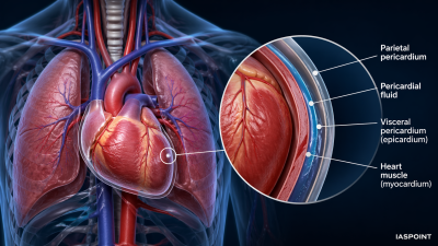
The human heart is a hollow, muscular organ derived from the mesoderm. It is located in the thoracic cavity within the mediastinum (the space between the lungs), slightly tilted toward the left. It is protected by a double-walled membranous sac called the pericardium.
- Pericardial Fluid: Found between the two layers of the pericardium, it acts as a lubricant to reduce friction during the continuous rhythmic contractions and protects the heart from mechanical shocks.
The Four-Chambered Architecture
The human heart is divided into four distinct chambers to ensure that oxygenated and deoxygenated blood do not mix, which supports high metabolic efficiency.
Atria (Receiving Chambers)
- These are the two upper, smaller chambers with thin muscular walls.
- Right Atrium: Receives deoxygenated blood from the entire body via the Superior Vena Cava (from upper body) and Inferior Vena Cava (from lower body).
- Left Atrium: Receives oxygenated blood from the lungs via four Pulmonary Veins.
Ventricles (Pumping Chambers)
- These are the two lower, larger chambers with thick muscular walls.
- Right Ventricle: Pumps deoxygenated blood to the lungs through the Pulmonary Artery.
- Left Ventricle: Pumps oxygenated blood to the entire body through the Aorta. It has the thickest walls of all chambers because it must generate enough pressure to circulate blood to the extremities.
The Valvular Apparatus
Valves are specialized structures that ensure blood flows in only one direction and prevent any backflow during contraction.
| Valve Type | Location | Function |
| Tricuspid Valve | Between Right Atrium and Right Ventricle | Prevents backflow into the Right Atrium. |
| Bicuspid (Mitral) Valve | Between Left Atrium and Left Ventricle | Prevents backflow into the Left Atrium. |
| Pulmonary Semilunar Valve | At the base of the Pulmonary Artery | Prevents backflow into the Right Ventricle. |
| Aortic Semilunar Valve | At the base of the Aorta | Prevents backflow into the Left Ventricle. |
Structural Support: Septa and Chordae Tendineae
- Interatrial Septum: A thin, muscular wall that separates the right and left atria.
- Interventricular Septum: A thick wall that separates the right and left ventricles.
- Chordae Tendineae: These are strong, fibrous “heartstrings” attached to the flaps of the tricuspid and bicuspid valves. They are anchored to Papillary Muscles in the ventricular walls, preventing the valves from folding backward into the atria during powerful ventricular contractions.
The Conducting System (Nodal Tissue)
The heart is myogenic, meaning the impulse for contraction originates within the heart’s own specialized muscle tissue rather than from external nerve stimulation.
- Sino-atrial Node (SAN): Located in the upper right corner of the right atrium. Known as the Pacemaker, it generates action potentials (70–75 per minute) that initiate the heartbeat.
- Atrio-ventricular Node (AVN): Located in the lower-left corner of the right atrium. It receives the impulse from the SAN and briefly delays it, ensuring the atria fully empty before the ventricles contract.
- Bundle of His & Purkinje Fibers: Specialized fibers that rapidly transmit the electrical impulse from the AVN throughout the ventricular walls, causing them to contract simultaneously.
Key Comparative Biology Facts
- Two-chambered Heart: Found in Fishes (1 Atrium, 1 Ventricle); utilizes single circulation.
- Three-chambered Heart: Found in Amphibians and Reptiles (2 Atria, 1 Ventricle); involves incomplete double circulation where blood mixes in the single ventricle.
- Four-chambered Heart: Found in Crocodiles, Birds, and Mammals; allows for complete separation of oxygenated and deoxygenated blood, essential for maintaining constant body temperature (Homeothermy).
Important Trivia for UPSC Prelims
- Mesodermal Origin: Like most muscles and the circulatory system, the heart develops from the middle embryonic layer (mesoderm).
- Heart Weight: In an average adult male, the heart weighs approximately 280–340 grams; in females, it is 230–280 grams.
- Coronary Arteries: The heart does not absorb nutrients from the blood passing through its chambers; it receives its own oxygenated supply via the left and right coronary arteries branching from the aorta.
- Fossa Ovalis: An oval depression in the interatrial septum, which is a remnant of the Foramen Ovale, an opening that exists in the fetal heart to bypass the lungs.