
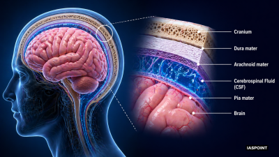
The human brain is the central information processing organ of the body, acting as the “command center.” It is protected by the cranium (skull) and is encased in three layers of connective tissue membranes called meninges: the outer Dura mater, middle Arachnoid mater, and inner Pia mater. The space between these layers is filled with Cerebrospinal Fluid (CSF), which provides buoyancy and acts as a shock absorber.
Major Divisions of the Brain
The brain is embryologically and functionally divided into three primary regions: the Forebrain, Midbrain, and Hindbrain.
1. Forebrain (Prosencephalon)
The largest and most complex part of the brain, responsible for higher-order functions.
- Cerebrum: Divided into two halves called Cerebral Hemispheres, connected by a dense tract of nerve fibers called the Corpus Callosum.
- Cerebral Cortex: The outer folded layer (Grey Matter) containing neuron cell bodies. The folds (gyri) and grooves (sulci) increase surface area for higher cognitive capacity.
- Lobes: Divided into Frontal (reasoning), Parietal (sensory), Temporal (hearing), and Occipital (vision) lobes.
- Thalamus: Acts as a major relay station for sensory and motor signaling.
- Hypothalamus: Located at the base of the thalamus. It regulates body temperature, urge for eating and drinking, and controls the secretion of pituitary hormones. It contains the osmocenters for water balance.
2. Midbrain (Mesencephalon)
Located between the thalamus/hypothalamus of the forebrain and the pons of the hindbrain.
- Cerebral Aqueduct: A canal passing through the midbrain.
- Corpora Quadrigemina: Four round swellings (lobes) on the dorsal portion involved in visual and auditory reflexes.
3. Hindbrain (Rhombencephalon)
Responsible for vital visceral functions and coordination.
- Pons: Consists of fiber tracts that interconnect different regions of the brain; it contains the pneumotaxic center that moderates breathing.
- Cerebellum: Known as the “little brain,” it has a very convoluted surface to accommodate more neurons. It coordinates muscular movements and maintains body equilibrium and posture.
- Medulla Oblongata: The lowest part of the brain that connects to the spinal cord. It contains centers controlling gastric secretions, cardiovascular reflexes, and respiration (rhythm center).
Functional Localization of the Brain
| Region | Primary Function |
| Frontal Lobe | Decision making, voluntary motor control, personality, and speech (Broca’s area). |
| Parietal Lobe | Processing somatosensory information (touch, pressure, pain). |
| Occipital Lobe | Visual processing and interpretation. |
| Temporal Lobe | Auditory perception, memory, and language comprehension (Wernicke’s area). |
| Limbic System | Called the “Emotional Brain”; includes the Amygdala and Hippocampus; regulates sexual behavior and emotions. |
The Brainstem
The brainstem is formed by the Midbrain, Pons, and Medulla Oblongata. It forms the connection between the brain and the spinal cord and is responsible for basic life-support functions like heartbeat and blood pressure. Note that the Cerebellum is NOT part of the brainstem.
Key Facts for UPSC Prelims
- Blood-Brain Barrier (BBB): A highly selective semipermeable border that protects the brain from circulating toxins and pathogens while allowing vital nutrients like glucose to pass.
- White Matter vs. Grey Matter: In the brain, Grey Matter (cell bodies) is on the outside (cortex), and White Matter (myelinated axons) is on the inside. This is reversed in the spinal cord.
- Ventricles: Four fluid-filled cavities within the brain where Cerebrospinal Fluid (CSF) is produced by the choroid plexus.
- Neuroplasticity: The brain’s ability to reorganize itself by forming new neural connections throughout life, essential for learning and recovering from injury.
- Hypothalamus-Pituitary Axis: The link between the nervous system and the endocrine system, crucial for maintaining internal homeostasis.