
The neuron is the fundamental structural and functional unit of the nervous system. It is a highly specialized cell designed to transmit information across the body via electrical and chemical signals.
Essential Components of a Neuron
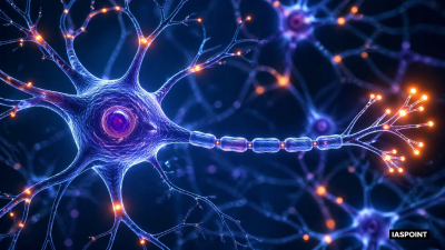
- Cell Body (Soma/Cyton): The metabolic center of the neuron containing the nucleus and various organelles. It contains Nissl’s granules, which are clusters of Rough Endoplasmic Reticulum (RER) and free ribosomes responsible for protein synthesis.
- Dendrites: These are short, highly branched fibers that project from the cell body. They act as the primary “input” terminals, receiving stimuli and conducting them toward the cell body.
- Axon: A long, slender projection that conducts impulses away from the cell body toward other neurons or effectors. The point where the axon joins the cell body is called the axon hillock, where the nerve impulse is typically generated.
- Myelin Sheath: A lipid-rich insulating layer found around many axons. In the Peripheral Nervous System (PNS), it is formed by Schwann cells; in the Central Nervous System (CNS), it is formed by Oligodendrocytes.
- Nodes of Ranvier: Periodic gaps in the myelin sheath along the axon. These gaps are essential for saltatory conduction, where the electrical signal “jumps” from node to node, significantly increasing the speed of transmission.
- Axon Terminals: The distal ends of the axon that branch out and end in bulb-like structures called synaptic knobs. These knobs contain synaptic vesicles filled with neurotransmitters.
Functioning of the Nervous System
The nervous system operates through a complex sequence of electrical excitation and chemical transmission.
1. Generation of Nerve Impulses (Action Potential)
- Resting Potential: When a neuron is not conducting an impulse, the axonal membrane is polarized. The outer side is positively charged while the inner side is negatively charged due to the distribution of Na^+ and K^+ ions maintained by the Sodium-Potassium Pump (which pumps 3 Na^+ out for every 2 K^+ in).
- Depolarization: When a stimulus reaches the threshold, Na^+ channels open, allowing Na^+ to rush into the cell. This reverses the polarity (inner side becomes positive), creating an Action Potential.
- Repolarization: K^+ channels then open and K^+ ions diffuse out, restoring the resting negative charge inside the membrane.
2. Transmission across Synapses
The junction between two neurons is called a synapse. Communication here is usually chemical.
- Synaptic Cleft: The fluid-filled space between the pre-synaptic neuron and the post-synaptic neuron.
- Neurotransmitter Release: When the action potential reaches the axon terminal, it triggers the influx of Ca2+ ions, causing synaptic vesicles to fuse with the membrane and release neurotransmitters (e.g., Acetylcholine, Noradrenaline) into the cleft.
- Binding: The neurotransmitters bind to specific receptors on the post-synaptic membrane, initiating a new electrical impulse in the next neuron.
Reflex Action and Reflex Arc
A reflex action is an immediate, involuntary response to a stimulus that does not involve conscious thought from the brain.
- Reflex Arc Pathway: Receptor (Sense organ) → Sensory Neuron (Afferent) → Spinal Cord (Interneuron) → Motor Neuron (Efferent) → Effector (Muscle/Gland).
- Significance: It provides a rapid survival mechanism to protect the body from harm (e.g., withdrawing a hand from a hot object).
Types of Nerve Fibers
| Type | Description | Occurrence |
| Myelinated | Wrapped in myelin sheath; high-speed transmission | Cranial and spinal nerves |
| Non-myelinated | Lacks myelin sheath; slower transmission | Autonomic and somatic neural systems |
Fact Sheet for UPSC Prelims
- Neuroplasticity: The ability of the nervous system to change its activity in response to intrinsic or extrinsic stimuli by reorganizing its structure, functions, or connections.
- Blood-Brain Barrier (BBB): A protective layer of endothelial cells and astrocytes that prevents most toxins and pathogens from entering the brain tissue.
- Glial-Neuron Ratio: While neurons transmit signals, glial cells are approximately 10 to 50 times more numerous and provide the necessary scaffolding and immune defense.
- Saltatory Conduction: This process in myelinated fibers is up to 100 times faster than continuous conduction in unmyelinated fibers.
- Synaptic Plasticity: The biological process where specific patterns of synaptic activity result in changes in synaptic strength; it is the biochemical basis for learning and memory.