
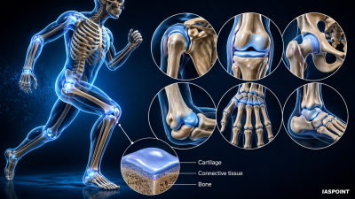
Cartilage is a resilient, smooth, and elastic type of connective tissue. It is a vital component of the musculoskeletal system, providing a framework for bone growth and ensuring frictionless movement at joints.
Key Structural Features
- Avascular Nature: Cartilage lacks blood vessels and nerves. It receives nutrients via diffusion from surrounding connective tissue (perichondrium) or synovial fluid. This explains why cartilage heals much slower than bone.
- Chondrocytes: The specialized cells of cartilage, which reside in small cavities called Lacunae.
- Extracellular Matrix (ECM): Composed of glycosaminoglycans, proteoglycans, collagen fibers, and sometimes elastin. The high water content in the matrix allows cartilage to be resilient and compressible.
- Perichondrium: A layer of dense irregular connective tissue that surrounds most cartilage (except at joints) and contains the blood supply.
Types of Cartilage
Cartilage is classified into three types based on the relative abundance of collagen and elastic fibers in the matrix.
| Type of Cartilage | Characteristics | Primary Locations |
| Hyaline Cartilage | Most abundant; bluish-white and glassy. Provides strong support with some flexibility. | Articular surfaces of long bones, Trachea, Bronchi, Larynx, and the Nose. |
| Elastic Cartilage | Contains numerous elastic fibers. Maintains shape while allowing great flexibility. | External Ear (Pinna) and the Epiglottis. |
| Fibrocartilage | Thick bundles of collagen; toughest type. Acts as a heavy-duty shock absorber. | Intervertebral discs, Pubic symphysis, and the Menisci of the knee. |
Functional Significance in the Human Body
- Template for Bone Growth: Most of the skeleton in a human fetus is initially made of hyaline cartilage, which is gradually replaced by bone through Endochondral Ossification.
- Shock Absorption: In the vertebral column and knee, fibrocartilage absorbs the impact of walking, running, and jumping.
- Friction Reduction: Articular cartilage (a type of hyaline) provides a smooth surface that allows bones to glide over each other with minimal wear.
- Structural Support: Provides permanent scaffolding for non-muscular structures like the ear and respiratory tubes.
Comparison: Cartilage vs. Bone
Understanding these differences is a frequent area of focus in Biology-based UPSC questions.
| Property | Cartilage | Bone |
| Primary Cells | Chondrocytes | Osteocytes |
| Matrix Type | Flexible, organic salts (Chondroitin). | Hard, inorganic salts (Calcium Phosphate). |
| Vascularity | Non-vascular (Avascular). | Highly vascular. |
| Growth Pattern | Appositional and Interstitial. | Primarily Appositional. |
| Nerve Supply | Absent. | Present. |
Important Facts and Trivia for Aspirants
- Epiphyseal Plate: This “growth plate” in children is made of hyaline cartilage. When it completely ossifies into bone, height growth stops.
- Costal Cartilage: The bars of hyaline cartilage that connect the ribs to the sternum, allowing the chest to expand during respiration.
- Ageing: As humans age, cartilage can undergo “calcification,” where it becomes hard and brittle, often confused with bone but lacking the organized structure of true osseous tissue.
- Articular Cartilage Loss: The destruction of hyaline cartilage at the joints is the primary cause of Osteoarthritis.
Related Clinical Conditions
- Chondritis: Inflammation of the cartilage.
- Herniated Disc: Occurs when the fibrocartilage of an intervertebral disc tears or bulges, often putting pressure on spinal nerves.
- Achondroplasia: A genetic disorder preventing the conversion of cartilage to bone, resulting in dwarfism.