
The large intestine is the terminal part of the alimentary canal. It is wider in diameter but shorter in length (approximately 1.5 meters) compared to the small intestine. Unlike the small intestine, it does not perform any significant chemical digestion of food. Its primary roles include the absorption of water, minerals, and certain vitamins, and the preparation of undigested waste for egestion.
Anatomical Structure
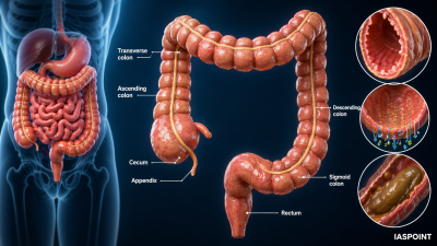
The large intestine is divided into three main regions: the Caecum, the Colon, and the Rectum.
1. Caecum
- Nature: A small, blind sac that represents the beginning of the large intestine.
- Symbiotic Micro-organisms: It hosts various symbiotic bacteria that aid in the synthesis of certain vitamins (like Vitamin K and B-complex).
- Vermiform Appendix: A narrow, finger-like tubular projection that is a vestigial organ in humans. It arises from the caecum and contains lymphoid tissue.
2. Colon
The colon is the longest part of the large intestine and is subdivided into four segments based on the direction of waste flow:
- Ascending Colon: Travels up the right side of the abdominal cavity.
- Transverse Colon: Crosses the abdomen from right to left.
- Descending Colon: Travels down the left side.
- Sigmoid Colon: An S-shaped segment that leads into the rectum.
3. Rectum and Anus
- Rectum: A muscular tube that serves as a temporary storage site for feces.
- Anus: The terminal opening of the alimentary canal. It is guarded by two sphincters: the Internal Anal Sphincter (involuntary smooth muscle) and the External Anal Sphincter (voluntary skeletal muscle).
Physiological Functions
The large intestine performs the following critical functions:
- Absorption: It absorbs nearly 90% of the water remaining in the chyme, along with minerals and some drugs.
- Mucus Secretion: It secretes mucus to lubricate the waste particles, helping them adhere together into feces and move smoothly toward the exit.
- Microbial Activity: The gut flora (microbiome) breaks down remaining carbohydrates (fermentation) and synthesizes Vitamin K, Biotin (B7), and Folic Acid (B9).
- Defecation: The elimination of solid waste, initiated by a reflex action (peristaltic waves in the colon).
Comparison: Small Intestine vs. Large Intestine
| Feature | Small Intestine | Large Intestine |
| Length | ~6–7 meters | ~1.5 meters |
| Diameter | Narrow | Wide |
| Villi | Present (for nutrient absorption) | Absent |
| Digestion | Primary site of chemical digestion | No significant digestive activity |
| Hormonal Role | Secretes CCK, Secretin, etc. | No major digestive hormones |
| Waste State | Chyme (liquid/semi-liquid) | Feces (solid/semi-solid) |
UPSC Prelims Trivia: The Gut Health
- Haustra: These are small pouches in the colon caused by the tonicity of the Taeniae Coli (three longitudinal bands of smooth muscle). They give the colon its segmented appearance.
- Ileocaecal Valve: A sphincter muscle situated at the junction of the ileum and the caecum. It prevents the backflow of fecal matter from the large intestine into the small intestine.
- Appendicitis: Inflammation of the vermiform appendix, often caused by a blockage, which can lead to rupture and infection of the abdominal cavity (peritonitis).
- Role in Immunity: The appendix and the Peyer’s patches in the nearby ileum contribute to the body’s mucosal immune system.
- Diarrhea vs. Constipation: Diarrhea occurs when the large intestine fails to absorb enough water (increased motility), whereas constipation occurs when too much water is absorbed due to slow movement.
Final Summary of the Alimentary Canal Flow
- Ingestion: Mouth/Buccal Cavity.
- Propulsion: Pharynx/Oesophagus.
- Mechanical/Initial Chemical Digestion: Stomach.
- Final Digestion & Absorption: Small Intestine.
- Water Recovery & Egestion: Large Intestine.