
The process of vision involves the conversion of light energy into electrical signals, a process known as Phototransduction.
The Optical Path of Light
To form a clear image, light must be focused precisely on the retina. This involves several structures acting as a refractive media.
- Refraction: The bending of light occurs primarily at the Cornea and subsequently through the Lens.
- Focusing: The Ciliary Muscles contract or relax to change the curvature of the lens, allowing the eye to focus on objects at different distances (Accommodation).
- Convergence: For near vision, both eyeballs turn slightly inward to ensure the light rays hit the fovea of both eyes.
Steps in Image Formation
- Entry of Light: Light reflected from an object enters the eye through the transparent cornea.
- Regulation: The Iris adjusts the size of the Pupil to control the intensity of light (constriction in bright light, dilation in dim light).
- Refraction and Inversion: The lens refracts the light such that it converges on the retina. The image formed on the retina is inverted (upside down) and real.
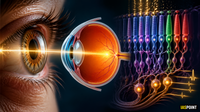
- Stimulation: Light reaches the photoreceptor cells (Rods and Cones) in the retina.
Biochemical Mechanism: Phototransduction
The retina contains light-sensitive proteins called Photopigments. In humans, these consist of Opsin (a protein) and Retinal (an aldehyde of Vitamin A).
- Dissociation: When light strikes the retina, it causes the Retinal to change shape and dissociate from Opsin.
- Conformational Change: This change in the structure of Opsin activates a G-protein called Transducin.
- Signal Generation: The activation leads to a change in the membrane permeability of the photoreceptor cells, resulting in a potential difference (action potential).
- Transmission: The signal is transmitted via Bipolar neurons to Ganglion cells, which converge to form the Optic Nerve.
- Interpretation: The Optic Nerve carries the impulse to the Visual Cortex in the occipital lobe of the brain, where the image is inverted back and perceived upright.
Comparison of Photoreceptor Functions
| Feature | Rods | Cones |
| Vision Type | Scotopic (Night/Dim light) | Photopic (Day/Bright light) |
| Acuity | Low resolution | High resolution (Visual Acuity) |
| Color Perception | Absent (Grayscale) | Present (Red, Green, Blue) |
| Distribution | Peripheral Retina | Concentrated in the Fovea Centralis |
| Pigment | Rhodopsin | Iodopsin |
Common Defects of Vision
Understanding these defects is critical for Science and Technology sections of the UPSC syllabus.
- Myopia (Near-sightedness): The eyeball is too long or the lens is too curved; the image forms in front of the retina. Corrected using Concave lenses.
- Hypermetropia (Far-sightedness): The eyeball is too short; the image forms behind the retina. Corrected using Convex lenses.
- Presbyopia: Age-related loss of lens elasticity, making it difficult to focus on near objects. Corrected with Bifocal lenses.
- Astigmatism: Caused by irregular curvature of the cornea or lens. Corrected using Cylindrical lenses.
Facts and Trivia for Prelims
- Persistence of Vision: The human eye retains an image for about 1/16th of a second. If images are flashed faster than this, they appear as continuous motion (the principle behind cinematography).
- Binocular Vision: Humans have two eyes positioned at the front of the head, allowing for Stereopsis (depth perception).
- Night Vision in Animals: Many nocturnal animals have a reflective layer behind the retina called the Tapetum Lucidum, which causes their eyes to “glow” in the dark and enhances night vision.
- Color Blindness: A genetic disorder (usually X-linked) where one or more types of cones are missing or non-functional. Red-Green color blindness is the most common.