
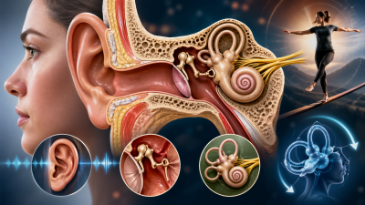
The ear is a complex vestibulo-cochlear organ responsible for two primary physiological functions: Hearing and Maintenance of Equilibrium (Balance). Anatomically, the ear is divided into three distinct sections: the Outer Ear, the Middle Ear, and the Inner Ear.
1. The Outer Ear
The outer ear is the entry point for sound waves and consists of structures that collect and direct sound.
- Pinna (Auricle): The visible external part made of elastic cartilage. It collects sound vibrations from the air.
- External Auditory Canal (Meatus): A tube leading inward that contains Ceruminous glands which secrete earwax (cerumen). This wax traps dust and repels insects.
- Tympanic Membrane (Eardrum): A thin, semi-transparent membrane that vibrates when struck by sound waves, marking the boundary between the outer and middle ear.
2. The Middle Ear
The middle ear is an air-filled cavity in the temporal bone that amplifies sound vibrations.
- Ear Ossicles: Three tiny bones connected in a chain-like fashion.
- Malleus (Hammer): Attached to the tympanic membrane.
- Incus (Anvil): The intermediate bone.
- Stapes (Stirrup): Attached to the Oval Window of the cochlea. It is the smallest bone in the human body.
- Eustachian Tube: A canal connecting the middle ear to the pharynx (throat). It equalizes air pressure on both sides of the eardrum, preventing it from rupturing during pressure changes (e.g., in an airplane).
3. The Inner Ear (Labyrinth)
The inner ear is a fluid-filled series of cavities known as the Labyrinth. It consists of two parts: the Bony Labyrinth and the Membranous Labyrinth.
- Perilymph and Endolymph: The bony labyrinth is filled with perilymph, while the membranous labyrinth (inside the bony one) is filled with endolymph.
The Cochlea (Organ of Hearing)
- Structure: A coiled, snail-like structure.
- Organ of Corti: Located on the basilar membrane, this is the actual sensory organ for hearing. It contains Hair Cells that act as auditory receptors.
- Mechanism: Vibrations from the stapes reach the perilymph, creating waves that move the hair cells. These cells generate nerve impulses sent via the Auditory Nerve to the brain.
The Vestibular Apparatus (Organ of Equilibrium)
Located above the cochlea, this system maintains body balance.
- Semicircular Canals: Three canals oriented in different planes to detect rotational movements of the head (Dynamic Equilibrium).
- Otolith Organs (Saccule and Utricle): Contain sensory hair cells and small calcium carbonate crystals (otoliths) to detect linear acceleration and head tilt (Static Equilibrium).
Functional Summary of Ear Components
| Component | Primary Function |
| Pinna | Collecting sound waves |
| Ear Ossicles | Amplifying sound vibration (approx. 20 times) |
| Eustachian Tube | Pressure equalization |
| Cochlea | Auditory transduction (Hearing) |
| Vestibular Apparatus | Equilibrium and Balance |
Key Facts for UPSC Prelims
- Sound Amplification: The middle ear ossicles act as a lever system to increase the force of sound vibrations before they enter the fluid-filled inner ear.
- Frequency Range: The human ear can typically perceive sound frequencies between 20 Hz and 20,000 Hz.
- Labyrinthitis: An inflammation of the inner ear that can cause both hearing loss and vertigo (dizziness/loss of balance).
- Decibel (dB): The unit used to measure sound intensity. Prolonged exposure to sounds above 85 dB can cause permanent damage to the hair cells in the Organ of Corti.