
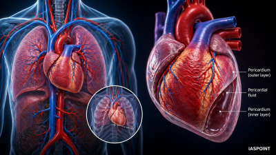
The human heart is a muscular organ derived from the mesoderm. It is situated in the thoracic cavity, in the space between the two lungs called the mediastinum, slightly tilted to the left. It is roughly the size of a closed fist and is protected by a double-walled membranous bag called the pericardium, which contains pericardial fluid to reduce friction during heartbeats.
Internal Structure and Chambers
The human heart is four-chambered, a characteristic shared with birds and crocodiles, which ensures the complete separation of oxygenated and deoxygenated blood.
- Atria (Auricles): The two upper, smaller chambers with relatively thin walls. They receive blood from the veins.
- Ventricles: The two lower, larger chambers. The walls of the ventricles are much thicker than the atria, with the left ventricle having the thickest wall as it must pump blood to the entire body.
- Septum: A muscular wall that prevents the mixing of blood. The inter-atrial septum separates the atria, while the inter-ventricular septum separates the ventricles.
The Valvular System
Valves are crucial for ensuring the unidirectional flow of blood and preventing backflow.
- Tricuspid Valve: Located between the right atrium and the right ventricle (has three muscular flaps).
- Bicuspid (Mitral) Valve: Located between the left atrium and the left ventricle (has two flaps).
- Semilunar Valves: Located at the base of the Pulmonary Artery (exiting the right ventricle) and the Aorta (exiting the left ventricle).
- Chordae Tendineae: Strong, fibrous strings (often called “heartstrings”) that connect the papillary muscles of the ventricles to the tricuspid and bicuspid valves to prevent them from everting during contraction.
The Nodal Tissue (Conducting System)
The human heart is myogenic, meaning the impulse for contraction is generated within the heart muscle itself by specialized nodal tissue.
- Sino-atrial Node (SAN): Located in the upper right corner of the right atrium. It is the Natural Pacemaker because it initiates the rhythmic contractile activity (70–75 times per minute).
- Atrio-ventricular Node (AVN): Located in the lower-left corner of the right atrium. it receives the impulse from the SAN and delays it slightly to allow atria to empty.
- Bundle of His: A bundle of nodal fibers that continues from the AVN and distributes the impulse to the ventricles.
- Purkinje Fibers: Fine fibers that branch throughout the ventricular musculature, causing the ventricles to contract.
The Path of Blood Flow
The heart functions as a dual pump facilitating Double Circulation.
| Side of Heart | Type of Blood | Source | Destination |
| Right Side | Deoxygenated | Superior & Inferior Vena Cava | Lungs (via Pulmonary Artery) |
| Left Side | Oxygenated | Pulmonary Veins | Body Tissues (via Aorta) |
Cardiac Output and Stroke Volume
- Stroke Volume: The volume of blood pumped out by each ventricle during a single cardiac cycle (approx. 70 ml).
- Heart Rate: The number of beats per minute (Average 72 bpm).
- Cardiac Output: The total volume of blood pumped by each ventricle per minute.
- Formula: Cardiac Output = Stroke Volume × Heart Rate
- For a healthy adult, this is approximately 5,000 ml (5 Liters) per minute.
Electrical Activity: The ECG
An Electrocardiogram (ECG) is a clinical tool used to record the electrical impulses generated by the heart.
- P-Wave: Represents the electrical excitation (depolarization) of the atria, leading to atrial contraction.
- QRS Complex: Represents the depolarization of the ventricles, which initiates ventricular contraction.
- T-Wave: Represents the return of the ventricles from excited to normal state (repolarization).
Essential Facts for UPSC Prelims
- Foramen Ovale: A hole in the septum between the two atria in a fetus, which normally closes at birth to become the Fossa Ovalis.
- Heart Sounds: * Lubb (First sound): Caused by the closure of the tricuspid and bicuspid valves at the start of ventricular systole.
- Dupp (Second sound): Caused by the closure of the semilunar valves at the start of ventricular diastole.
- Coronary Circulation: The heart muscle receives its own blood supply via the Coronary Arteries. A blockage here leads to a Heart Attack (Myocardial Infarction).
- Artificial Pacemaker: A medical device implanted when the SAN fails to function, used to stimulate the heart with electrical impulses.