
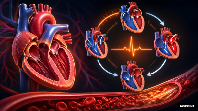
The cardiac cycle refers to the sequential events that occur in the heart from the beginning of one heartbeat to the beginning of the next. It involves a coordinated series of electrical and mechanical events including contraction (systole) and relaxation (diastole) of the heart chambers.
- Duration: In a healthy adult with a heart rate of 72 beats per minute, one cardiac cycle lasts approximately 0.8 seconds.
- Significance: It ensures that blood is pressurized and circulated efficiently throughout the pulmonary and systemic circuits.
Phases of the Cardiac Cycle
The 0.8-second cycle is divided into three primary physiological phases.
1. Joint Diastole (0.4 seconds)
In this initial phase, all four chambers of the heart are in a relaxed state.
- Valves: The tricuspid and bicuspid (mitral) valves are open, while the semilunar valves (aortic and pulmonary) are closed.
- Blood Flow: Blood from the pulmonary veins and vena cava flows into the left and right atria, and then directly into the respective ventricles.
- Filling: Approximately 70% of ventricular filling occurs passively during this phase.
2. Atrial Systole (0.1 seconds)
The Sino-atrial Node (SAN) generates an action potential, causing both atria to contract simultaneously.
- Function: This contraction increases the flow of blood into the ventricles by about 30%.
- End Result: At the end of this phase, the ventricles are filled with their maximum volume of blood, known as the End Diastolic Volume (EDV).
3. Ventricular Systole (0.3 seconds)
The action potential is conducted to the ventricles via the AV Node and Bundle of His, causing ventricular contraction.
- AV Valve Closure: As ventricular pressure rises, the bicuspid and tricuspid valves close to prevent backflow into the atria. This produces the first heart sound (“Lubb”).
- Semilunar Valve Opening: When ventricular pressure exceeds the pressure in the aorta and pulmonary artery, the semilunar valves open.
- Ejection: Blood is pumped into the circulatory pathways. The volume of blood pumped out by each ventricle is the Stroke Volume (approx. 70 ml).
Ventricular Diastole and the Second Heart Sound
Following systole, the ventricles relax (Ventricular Diastole).
- Semilunar Valve Closure: As ventricular pressure falls, blood in the great arteries tries to flow back, causing the semilunar valves to snap shut. This produces the second heart sound (“Dupp”).
- Cycle Reset: As pressure continues to drop, the AV valves open again, and the heart returns to the state of Joint Diastole.
Summary Table: Events in a Single Cardiac Cycle
| Phase | Duration | Status of Atria | Status of Ventricles | Status of AV Valves | Status of Semilunar Valves |
| Joint Diastole | 0.4s | Relaxed | Relaxed | Open | Closed |
| Atrial Systole | 0.1s | Contracting | Relaxed | Open | Closed |
| Ventricular Systole | 0.3s | Relaxed | Contracting | Closed | Open |
Cardiac Volume Terminology
- End Diastolic Volume (EDV): The volume of blood in a ventricle at the end of filling (approx. 120 ml).
- End Systolic Volume (ESV): The volume of blood remaining in a ventricle after contraction (approx. 50 ml).
- Stroke Volume (SV): The amount of blood ejected per beat (EDV – ESV = 70 ml).
- Cardiac Output: The total volume of blood pumped per minute (Stroke Volume × Heart Rate ≈ 5 Liters).
Clinical Correlation: Heart Sounds
The heart sounds are of significant clinical diagnostic value and are heard using a Stethoscope.
- Lubb (S1): Low-pitched, louder, and of longer duration. Associated with the closure of Atrioventricular (AV) valves.
- Dupp (S2): Higher-pitched, shorter, and sharper. Associated with the closure of Semilunar valves.
- Murmurs: Abnormal sounds heard when valves are defective, causing turbulent blood flow or regurgitation.
Important Facts for UPSC Prelims
- Atrial Diastole: While the ventricles are in systole (0.3s) and the heart is in joint diastole (0.4s), the atria are in a state of diastole for a total of 0.7 seconds.
- Pulse: The rhythmic expansion of an artery as blood is ejected from the left ventricle. Pulse rate is usually equal to heart rate.
- Regulation: During exercise, the duration of the cardiac cycle shortens (specifically the diastole phase) to accommodate a higher heart rate and increased cardiac output.
- Isovolumetric Contraction: A brief period at the start of ventricular systole where all valves are closed and pressure builds without a change in volume.