
Breathing is the physical process of moving air into and out of the lungs to facilitate gas exchange. It operates on the principle of Boyle’s Law, which states that the pressure of a gas is inversely proportional to its volume at a constant temperature.
Pressure Gradients
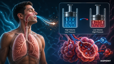
Breathing depends on the creation of a pressure gradient between the atmosphere and the alveoli.
- Inspiration: Occurs when the intra-pulmonary pressure is lower than the atmospheric pressure (negative pressure in the lungs).
- Expiration: Occurs when the intra-pulmonary pressure is higher than the atmospheric pressure.
Muscles Involved in Respiration
The movement of air is facilitated by a specialized set of muscles that alter the volume of the thoracic cavity.
- Diaphragm: A dome-shaped muscle that forms the floor of the thoracic cavity. Its contraction increases the vertical (antero-posterior) volume.
- External Intercostal Muscles: Located between the ribs. Their contraction lifts the ribs and the sternum, increasing the volume in the dorso-ventral axis.
- Internal Intercostal Muscles: Primarily used during forceful expiration to pull the ribs downward.
The Process of Inspiration (Active Process)
Inspiration is an active phase requiring muscular contraction and energy (ATP).
- Muscle Contraction: The diaphragm contracts and flattens; external intercostal muscles contract, pulling the rib cage up and out.
- Volume Increase: The overall volume of the thoracic chamber increases.
- Pressure Drop: As the thoracic volume increases, the intra-pulmonary pressure drops below atmospheric pressure.
- Air Inflow: Air rushes from the higher atmospheric pressure into the lungs to equalize the pressure.
The Process of Expiration (Passive Process)
Normal expiration is generally passive, relying on the elastic recoil of the lungs and relaxation of muscles.
- Muscle Relaxation: The diaphragm and external intercostal muscles relax, returning to their original positions.
- Volume Decrease: The thoracic volume decreases, which in turn reduces the pulmonary volume.
- Pressure Rise: The intra-pulmonary pressure increases slightly above atmospheric pressure.
- Air Outflow: Air is expelled from the lungs.
Forced Breathing
During vigorous exercise or respiratory distress, additional muscles (abdominal muscles and internal intercostals) are recruited. This is an active process for both inspiration and expiration.
Respiratory Volumes and Capacities
Understanding these values is crucial for diagnosing pulmonary health in UPSC-related science questions.
| Term | Definition | Average Value |
| Tidal Volume (TV) | Air inspired/expired during normal breathing. | 500 mL |
| Inspiratory Reserve Volume (IRV) | Extra air inspired forcibly after normal inspiration. | 2500–3000 mL |
| Expiratory Reserve Volume (ERV) | Extra air expired forcibly after normal expiration. | 1000–1100 mL |
| Residual Volume (RV) | Air remaining in lungs after forced expiration. | 1100–1200 mL |
| Vital Capacity (VC) | Total exchangeable air (TV + IRV + ERV). | 3500–4500 mL |
Regulation of Breathing
The body maintains a constant rhythm of breathing through neural and chemical controls.
- Respiratory Rhythm Center: Located in the Medulla Oblongata; it is the primary center for regulating the rhythm.
- Pneumotaxic Center: Located in the Pons region of the brain; it can moderate the functions of the rhythm center by reducing the duration of inspiration.
- Chemosensitive Area: Situated adjacent to the rhythm center, it is highly sensitive to CO2 and hydrogen ions. An increase in these substances triggers the center to eliminate them.
- Peripheral Chemoreceptors: Located in the Aortic arch and Carotid artery; they also sense CO2 and H^+ concentration changes and send signals to the Medulla.
Important Trivia
- Spirometer: An instrument used to measure the volume of air inspired and expired for clinical assessment of pulmonary functions.
- Role of Oxygen: Surprisingly, the role of oxygen in the regulation of respiratory rhythm is quite insignificant compared to CO2 and H^+ levels.
- Surface Tension: Alveoli are coated with surfactant, which prevents them from collapsing during expiration by reducing surface tension.